History of Presenting Condition
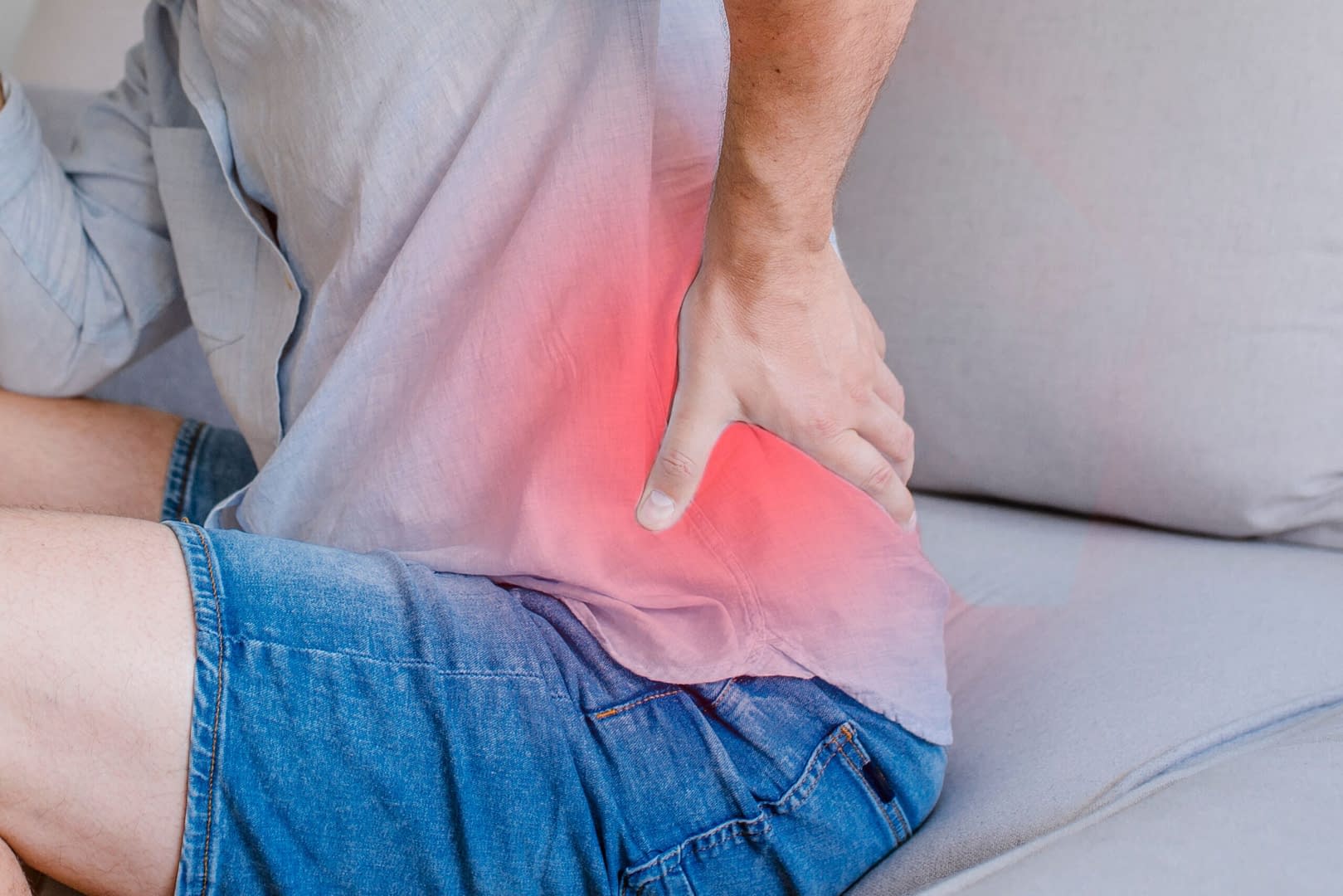
A 78-year-old male presents with left-sided lower back pain that radiates from the pelvis to the front of his thigh and just below the knee. This pain began two weeks ago while he was sailing. At the time, he used his left foot to operate a switch, but the movement combined with the boat’s rocking led him to feel weakness in his left leg.
The patient reports that his left knee has been unstable, causing his leg to “give way” between six and ten times while walking. He denies any numbness, tingling, or other neurological symptoms. He has experienced minor bladder leaks due to not reaching the bathroom in time, but denies saddle anaesthesia or any symptoms suggesting cauda equina syndrome (CES), which have been ruled out.
He rates his pain as 9 out of 10 on the Numeric Pain Rating Scale (NPRS). He arrived for the consultation using a walking stick and was visibly in extreme pain, unable to sit down in a chair.
SH: The patient has been an avid sailor, typically sailing once a week for many years. He lives alone in a bungalow.
MH: The patient is pre-diabetic but has no other known comorbidities. He has been taking paracetamol regularly, but reports no relief from this medication.
ICE: The patient expected to receive an MRI scan of his lumbar spine to investigate his symptoms further.
Assessment
- Antalgic very slow gait with a stick
- In sitting, had weak and painful L2/3, S2 myotomes – 4/5, dermatomes – intact
- Hip flexion in sitting painful and restricted range
- Reflex equal b/l
- SLUMP –ve, stretch pain over hams
- Knee ligaments intact, ant/post drawer test -ve
- Had tenderness over lx spine globally, lx paraspinals tightness/ tenderness L>R, tightness over L quads
Differential Diagnosis and Reasoning
The differential diagnosis included femoral radicular pain, hip pain, osteoarthritis (OA), and possible stress fracture. Spinal red flags, including cauda equina syndrome, metastatic spinal disease, and inflammatory conditions, were deducted at this stage.
A shared decision-making process with the patient determined that an MRI was not indicated at this stage for lumbar spine.
The patient was encouraged to engage in gentle exercises and use OTC NSAIDs after ruling out any contradictions/ allergies.
Follow-up booked in three weeks which indicated localised pain over L hip/ groin area with FABER/FADDIR+, morning stiffness+, abductor weakness, unable to straighten knee in supine, suggesting potential articular joint involvement?
An X-ray of the left hip was ordered to evaluate for OA or a stress fracture. The imaging showed severe hip OA with complete joint space loss and a femoral cam deformity.
The patient has been referred to the orthopaedic triage team for further management and discussion of surgical options.
Summary
- Importance of differentiating between radicular pain and radiculopathy.
- Imaging does not often change the initial management and outcomes of patients with back pain because the reported imaging findings are usually common and not necessarily related to the individual’s symptoms.
- The SLUMP test and NPRS are effective tools for assessing neuropathic symptoms, e.g. asking the patient to perform a seated straight leg raise (SLR) to check function of quadriceps.
- The SLUMP test has sensitivity which ranges from 44% – 87% and specificity ranges from 23 to 63%.
- Subjective findings, such as understanding the mechanism of injury, family history, pain referral to the groin or buttocks, reduced walking capacity, and morning stiffness, are useful in confirming a diagnosis of osteoarthritis.
- A cluster of objective findings, including pain during squats, referred groin pain on passive abduction/adduction, abduction weakness, and restricted adduction/internal rotation, has a positive likelihood ratio of 15.4.
- OTC medication was recommended to help reduce load over GPs.
- Patient education included information on available referral pathways and local services.


