Iliotibial Band Syndrome
1. Introduction
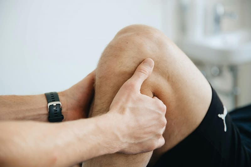
Iliotibial band syndrome is a condition that usually presents as pain on the outside of the knee. There is debate about the exact nature of the condition. Previously we believed that it was a friction syndrome, caused by rubbing over the bony part of the outside of the knee. More recently, however, we now understand that it is more of an impinging of the tissue at the side of the knee (1). This is important as it changes how we look to manage and ultimately resolve, the condition.
Frequently Asked Questions
- Iliotibial band syndrome is a condition that usually presents as pain on the outside of the knee. It normally occurs because of overload due to prolonged or repeated bouts of exercise.
- Iliotibial band syndrome is not common in the general population (1).
- It does make up about 10% of injuries suffered by runners (2).
- It can also occur in other sports and physical activity although less commonly.
- No.
- With the right rehabilitation approach, iliotibial band syndrome generally recovers well without the need for further intervention (4).
- It is not linked to other serious pathology.
- This condition is predominantly something that occurs in runners.
- It can occur in those involved in all running-based sports.
- It is more common in females (3, 7).
- Pain on the side of the knee.
- Pain is typically felt when loading the knee (walking/running).
- If symptoms are significant, pain and stiffness may be present after long periods of inactivity.
- Typically, pain will increase following a period of exercise (2).
- Reduce the volume and intensity of your exercise (2).
- Increase the flexibility of the muscles around the area.
- Improve the strength/stability of the hip (5).
- In some cases, a physiotherapist may assess your foot posture and recommend insoles.
- Advice by a qualified physiotherapist will be helpful in most cases.
- This will depend upon several factors including, but not limited to: the duration of symptoms, age, adherence to rehabilitation, amount of training you are attempting to complete.
- We would expect to see improvement in symptoms within 2-3 months and certainly no longer than 3-4 months (2).
We recommend consulting a musculoskeletal physiotherapist to ensure exercises are best suited to your recovery. If you are carrying out an exercise regime without consulting a healthcare professional, you do so at your own risk.
2. Signs and Symptoms
The typical signs and symptoms of the condition are a sharp pain on the outside of the knee, particularly when the foot hits the floor (walking or running). There is unlikely to be any swelling and it is uncommon to have any ‘giving way’, locking or clicking with this condition. Some people will describe the pain as a throbbing or burning pain (5).
Most commonly pain is the main complaint, with stiffness becoming progressively more of a problem, particularly after periods of inactivity.
3. Causes
There are a number of possible causes of the condition but the most common are;
- Hip muscle weakness – weakness of the hip muscles can increase the stress on the outside of the knee, particularly when you are fatigued from exercise (4).
- Excessive training load – training load that your body is not accustomed to, or training that involves a significant amount of volume without sufficient rest, can cause a number of overuse conditions, in particular iliotibial band syndrome (7).
- Poor running technique – typically running with what is termed a crossover gait can increase the load to the iliotibial band.
- Poor foot mechanics – excessive or poor control of pronation (the way your foot and arches flatten to distribute force when landing) of the foot can also increase the load on the structure due to an increase of internal rotation of the tibia.
4. Risk Factors
This is not an exhaustive list. These factors could increase the likelihood of someone developing iliotibial band syndrome. It does not mean everyone with these risk factors will develop symptoms.
- History of previous injuries (age < 34 years).
- Tight lateral fascial band (also known as the ITB).
- Improper footwear.
- Training factors such as running surface, high weekly mileage, lack of recovery, downhill running.
- Leg length differences.
- Increased angle of flexion of the knee at heel strike.
- Muscular weakness of knee extensors, knee flexors and hip abductors (5).

5. Prevalence
Iliotibial band syndrome is very rarely seen in the general population. It is one of the most common injuries in runners presenting with lateral knee pain, with an incidence estimated to be between 5% and 14% (2). Further studies indicate that iliotibial band syndrome is responsible for approximately 22% of all lower extremity injuries (9).
6. Assessment & Diagnosis
A musculoskeletal physiotherapist can provide you with an accurate and timely diagnosis by obtaining a detailed history of your symptoms. A series of physical tests might be performed as part of your assessment to rule out other potentially involved structures and gain a greater understanding of your physical abilities. Any tests we do will be considered alongside your symptoms to ensure we have an accurate working diagnosis
Your physiotherapist will want to know how your condition affects you day-to-day so that treatment can be tailored to your needs and personalised goals can be established. Intermittent reassessment will ascertain if you are making progress towards your goals and will allow appropriate adjustments to your treatment to be made.
It is rarely necessary in this condition to require any scans or X-rays to be done immediately as physiotherapy will most likely be the first line of treatment. If symptoms persist or get worse then an MRI scan may be used to understand what else could be causing the symptoms.
7. Self-Management
As part of the sessions with your physiotherapist, they will help you to understand your condition and what you need to do to help the recovery from your iliotibial band syndrome. This may include reducing the amount or type of activity, as well as other advice aimed at reducing your pain. It is important that you try and complete the exercises you are provided as regularly as possible to help with your recovery. Rehabilitation exercises are not always a quick fix, but if done consistently over weeks and months then they will, in most cases, make a significant difference.
8. Rehabilitation
It is widely agreed that the best starting point in the management of iliotibial band syndrome is physiotherapy-led rehabilitation (2, 7, 8). This will focus on optimising the flexibility and strength of the hip and foot.
9. Iliotibial Band Syndrome
Rehabilitation Plans
Our team of expert musculoskeletal physiotherapist have created rehabilitation plans to enable people to manage their condition. If you have any questions or concerns about a condition, we recommend you book an consultation with one of our clinicians.
What Is the Pain Scale?
The pain scale or what some physios would call the Visual Analogue Scale (VAS), is a scale that is used to try and understand the level of pain that someone is in. The scale is intended as something that you would rate yourself on a scale of 0-10 with 0 = no pain, 10 = worst pain imaginable. You can learn more about what is pain and the pain scale here.
This programme focuses on better recruitment of the gluteal muscles, as well as increasing the flexibility of the thigh muscles and hip rotators. It must be understood that whilst pain during and after these exercises cannot be totally avoided, a significant increase in pain is not desirable and can in fact hinder progress. Pain should not exceed 3/10 on your perceived pain scale.
- 0
- 1
- 2
- 3
- 4
- 5
- 6
- 7
- 8
- 910
More focus is given to the strength of the hip muscles and single-leg stability. Again, it must be understood that whilst pain during and after these exercises cannot be totally avoided, a significant increase in pain is not desirable and can in fact hinder progress. Pain should not exceed 4/10 on your perceived pain scale.
- 0
- 1
- 2
- 3
- 4
- 5
- 6
- 7
- 8
- 910
This programme is a further progression with exercises that challenge the strength of the area in a way that prepares you for a normal level of training. Pain should not exceed 3/10 on your perceived pain scale.
- 0
- 1
- 2
- 3
- 4
- 5
- 6
- 7
- 8
- 910
10. Return to Sport / Normal life
This condition is common in runners but the success rate of treatment through physiotherapy is very high. In most cases, it will be possible to return to a normal level of activity on completion of a course of physiotherapy.
11. Other Treatment Options
Typically, exercise is the best strategy for dealing with iliotibial band syndrome. In the past, it was believed that techniques such as massage and ultrasound to the iliotibial band itself were effective. However, recent research brings this assumption into question.
Local use of radial shockwave therapy (RSWT) is believed to stimulate the healing of soft tissue and reduce the activation of pain receptors. Radial shockwave therapy has been shown to be effective as part of a rehabilitation programme for runners with iliotibial band syndrome (8).
References
- Geisler, Paul & Lazenby, Todd. (2016). Iliotibial Band Impingement Syndrome: An Evidence-Informed Clinical Paradigm Change. International Journal of Athletic Therapy and Training. 22. 1-30. 10.1123/ijatt.2016-0075.
- Baker RL, Fredericson M. Iliotibial Band Syndrome in Runners: Biomechanical Implications and Exercise Interventions. Physical médicine and réhabilitation clinics of North America, 2016; 27(1):53-77
- Christopher D. Stickley, Melanie M. Presuto, Kara N. Radzak, Christina M. Bourbeau, Ronald K. Hetzler; Dynamic Varus and the Development of Iliotibial Band Syndrome. J Athl Train 1 February 2018; 53 (2): 128–134.
- Robert L. Baker, Richard B. Souza, Mitchell J. Rauh, Michael Fredericson, Michael D. Rosenthal, Differences in Knee and Hip Adduction and Hip Muscle Activation in Runners With and Without Iliotibial Band Syndrome, PM&R, Volume 10, Issue 10, 2018, Pages 1032-1039,
- Peixin Shen, Dewei Mao, Cui Zhang, Wei Sun & Qipeng Song (2019) Effects of running biomechanics on the occurrence of iliotibial band syndrome in male runners during an eight-week running programme—a prospective study, Sports Biomechanics, DOI: 10.1080/14763141.2019.1584235
- McKay, J., Maffulli, N., Aicale, R. and Taunton, J., 2020. Iliotibial band syndrome rehabilitation in female runners: a pilot randomized study. Journal of Orthopaedic Surgery and Research, 15, pp.1-8.
- Brown, A.M., Zifchock, R.A., Lenhoff, M., Song, J. and Hillstrom, H.J., 2019. Hip muscle response to a fatiguing run in females with iliotibial band syndrome. Human movement science, 64, pp.181-190.
- Weckström K, Söderström J. Radial extracorporeal shockwave therapy compared with manual therapy in runners with iliotibial band syndrome, Journal of Back and Musculoskeletal Rehabilitation, 2016; 29(1):161-70. doi: 10.3233/BMR-150612.
- Lavine R. Iliotibial band friction syndrome. Current Reviews in Musculoskeletal Medicine, 2010; 3(1-4) :18–22
Other Conditions in
Hips & Pelvis, Upper Legs, Knees
Sacroiliac Joint Dysfunction
Pain originating from the sacroiliac joint at the base of your back where the spine joins the pelvis.
Quadriceps Tendinopathy
The quadriceps are a group of four muscles found on the anterior thigh. They work together to extend, straighten, the knee, and control knee flexion when you are on your feet. They play an important role in activities such as running, standing up from a chair, climbing stairs, jumping, squatting and kicking.
Proximal Hamstring Tendinopathy
Pain and weakness under the buttock or the back of your upper thigh caused by tendon issues.
Pelvic Girdle Pain
Typically seen in pregnancy causing pain, instability and limitation of mobility and functioning of the pelvic joints.
Pelvic Floor Dysfunction
The inability to effectively control the muscles of your pelvic floor, leading to issues with continence and pain.
Meralgia Paraesthetica
Pain on the outside thigh caused by compression and inflammation of the nerve that supplies that area
Lumbar Disc Injury
Lumbar discs sit between each of the bones of the spine. Problems can occur when these discs become irritated.
Low Back Pain and Sciatica
Sciatica is a symptom describing pain and/or pins and needles down the back of the leg.
Hip Replacement Surgery
Replacement of the hip ball and socket joint, typically as a result of severe osteoarthritis or trauma.
Hip Osteoarthritis
Common age-related changes to the structure of the hip joint may be associated with pain, stiffness and loss of function.
Hamstring Strain/Tear
Hamstring strain injuries are an over-stretch or tear to one or more of the muscles located at the back of the thigh.
Greater Trochanteric Pain Syndrome
A condition affecting the tendons that insert into outside of the hip. A common cause of pain felt around the hip and pelvis.
Femoroacetabular Impingement
A condition that results in pain in the groin, hip and down the front of the thigh.
Femoral Nerve Radiculopathy
This is where the nerve that supplies the front of the leg is irritated and causes pain/numbess.
Coccydynia
Coccydynia is the medical term used to describe pain in your coccyx (tail bone).
Benign Joint Hypermobility Syndrome
Common age related changes to the structure of the knee joint which may be associated with pain, stiffness and loss of function.
Adolescent Hip Dysplasia
A result of an abnormality of the hip joint anatomy resulting in pain in the hip with occasional instability.
Adductor-Related Groin Pain
Localised discomfort to the inner upper thigh and groin.